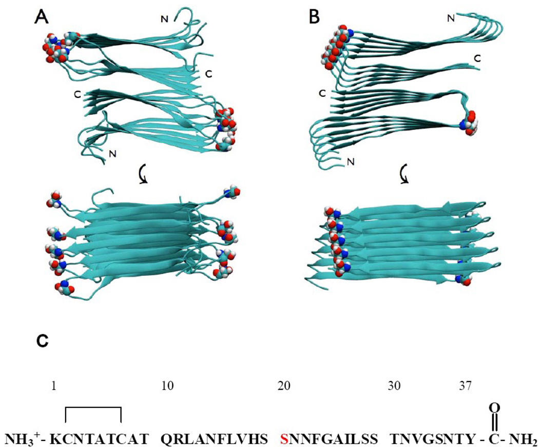
Figure 1.
Structural models of the wild type IAPP amyloid fiber showing the location of Ser-20. Two views are shown: a top down view and an image rotated by 90 degrees. Ser-20 is shown in space-filling representation. (A) The model developed by the NIH group. (B) The model developed by the UCLA group. (C) The primary sequence of human IAPP, residue Ser-20 is colored in red. The wild type peptide contains a disulfide bridge between Cys-2 and Cys-7 and has an amidated C-terminus.

