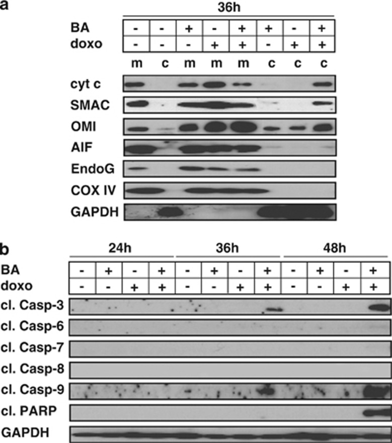
Figure 2.
Release of apoptogenetic factors and activation of caspases during synergistic apoptosis induction. (a) JURKAT cells were stimulated with doxo and BA for 36 h and fractionated investigation of cytosolic (c) and mitochondrial (m) extracts was performed by western blot. GAPDH and COX IV served as loading controls. For the clearness of presentation, the order of samples from the original blot was rearranged without any further modifications. (b) JURKAT cells were stimulated as in Figure 2a for time periods indicated and western blot analysis was performed of total cellular extracts. For the detection of all caspase-cleavage products, TRAIL-stimulated cells were used as positive control (data not shown). Experimental procedure and drug concentrations were applied as in Figure 1a

