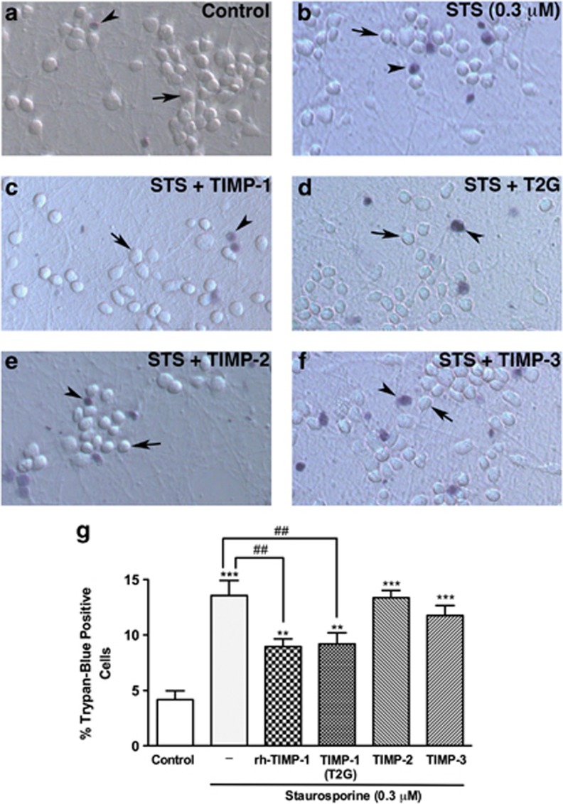
Figure 3.
TIMP-1-mediated neuroprotection is specific to TIMP-1 and MMP-independent. The cell viability assay was performed in human neurons after exposure of STS (0.3 μM) for 18 h with or without 50 ng/ml of TIMP-1, -2, -3 or 28 ng/ml T2G. Panels are representative micrographs of 10 random fields (original magnification × 200) from two replicates of control (a), STS (b) and STS with TIMP-1 (c), T2G (d), TIMP-2 (e) or TIMP-3 (f). Trypan-blue positive cells were considered as dead cell (represented by arrowhead) while, cells that excluded trypan-blue were counted as live (represented by arrow) and percentage of trypan-blue positive cells was calculated (g). Data is representative of at least three independent biological replicates and presented as mean±S.E.M. Symbols centered over the error bar indicate the relative level of the significance compared with untreated control (**P<0.01 and ***P<0.001) or STS-alone (♯♯P<0.01)

