Figure 1.
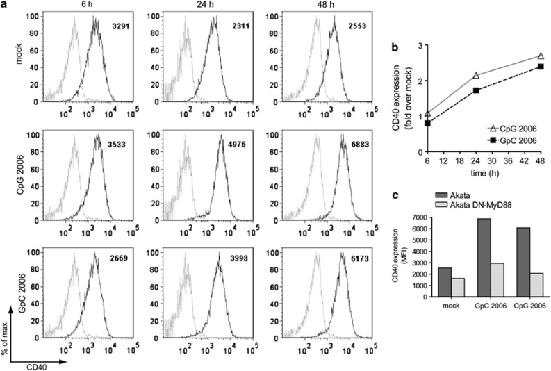
TLR9 agonists induce CD40 expression in Akata cells in a MyD88-dependent manner. Akata cells or DN-MyD88 Akata cells were left untreated or treated with 0.5 μM ODN CpG-2006 or ODN GpC-2006 for 48 h. After 6, 24 and 48 h, 105 cells were harvested, washed with PBS and stained with PE-Cy5-conjugated mouse anti-human CD40 antibody or a PE-Cy5 mouse IgG1κ isotype control and analyzed with a flow cytometer. (a) FACS plots of Akata cells. Isotype control: shaded gray line; anti-CD40 PE-Cy5: black line. Numbers indicate the mean fluorescence intensity (MFI) (b) Quantification of (a). (c) Comparison of CD40 expression in Akata cells and DN-MyD88 Akata cells

