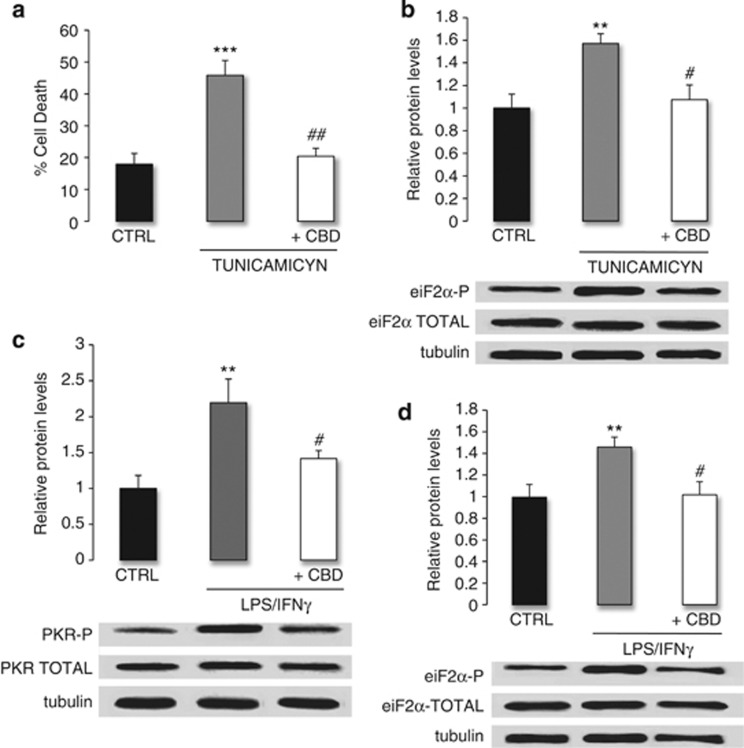
Figure 4.
OPC death is mediated by ER stress, an effect that is attenuated by CBD through decreased PKR and eiF2α phosphorylation in conditions of inflammation. (a) CBD attenuated tunicamycin-induced OPC death. OPCs were incubated with tunicamycin (1 μg/ml) in the presence or absence of CBD (1 μM), and cell death was quantified 24 h later by the LDH method. The data represent the mean±S.E.M. of three independent cultures analyzed in triplicate, and the statistical significance was determined using Kruskal–Wallis ANOVA followed by Mann–Whitney U test: ***P⩽0.001 versus untreated cells, ##P⩽0.01 versus cells exposed to tunicamycin alone. (b) Tunicamycin treatment induced the eiF2α phosphorylation, an effect that was attenuated by CBD. OPCs were incubated with tunicamycin (1 μg/ml) in the presence or absence of CBD (1 μM). Total protein extracts were prepared 5 min later and the phosphorylated (38 kDa) and total (38 kDa) eiF2α was assessed in western blots probed with specific antibodies. The data represent the mean±S.E.M. optical density normalized to tubulin from four independent cultures analyzed in triplicate, and the statistical significance was determined using Kruskal–Wallis ANOVA followed by Mann–Whitney U test: **P⩽0.01 versus untreated cells, #P⩽0.05 versus cells exposed to tunicamycin alone. (c and d) Inflammation-induced PKR and eiF2α phosphorylation, an effect that was attenuated by CBD. OPCs were treated with LPS/IFNγ in the presence or absence of CBD (1 μM). Total protein extracts were prepared 5 min later and PKR (phosphorylated, 68 kDa; total, 68 kDa) and eiF2α (phosphorylated, 38 kDa; total, 38 kDa) was assessed in western blots probed with specific antibodies. The data represent the mean±S.E.M. optical density normalized to tubulin from five cultures, and the statistical significance was determined using Kruskal–Wallis ANOVA followed by Mann–Whitney U test: **P⩽0.01 versus untreated cells, #P⩽0.05 versus cells exposed to LPS/IFNγ alone

