
Figure 2.
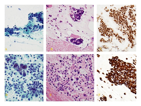
Distinct bladder wall and rectal wall FNA sites revealing high-grade urothelial carcinoma and adenocarcinoma, respectively, synchronously in the same patient. (A) Rectal wall: positive for malignancy. Adenocarcinoma consistent with colorectal primary. (Pap Stain); (B) rectal wall: positive for malignancy. Adenocarcinoma consistent with colorectal primary (H&E stain); (C) rectal wall: neoplastic cells are positive for CDX2 immunostaining; (D) bladder wall: positive for malignancy. High-grade urothelial carcinoma. (Pap Stain); (E) bladder wall: positive for malignancy. High-grade urothelial carcinoma. (H&E stain); (F) bladder wall: CK903 positive neoplastic cells with immunostaining.