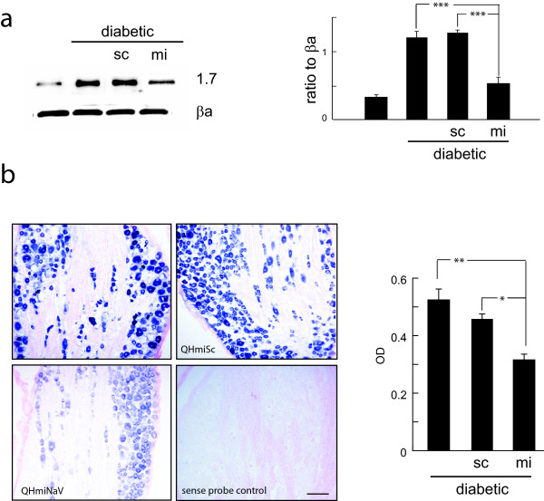
Figure 3.
a. NaV1.7 levels in DRG of diabetic animals inoculated with QHmiNaV demonstrates reduction in protein compared to QHmiSc 4 weeks after inoculation (*** p < 0.005). Data presented as ratio to β-actin (βa). b. In situ hybridization using a probe specific for NaV1.7 in DRG from diabetic animals, without treatment or inoculated with QHmiSc or QHmiNaV as indicated. A sense probe showed no staining. The average optical density of DRG neurons in each condition was determined using a PC based image analysis program (MCID). * p < 0.05; ** p < 0.01.

