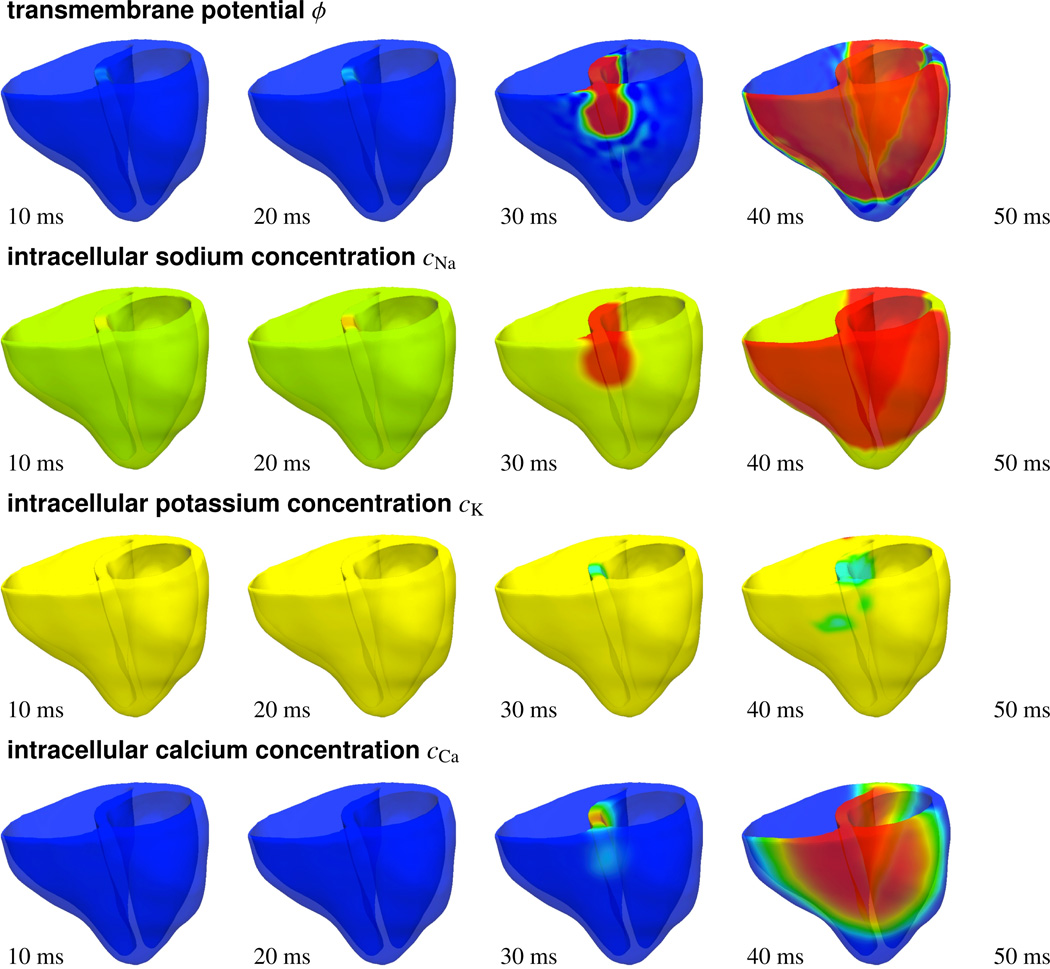
Figure 11.
Photostimulation of a human heart. Spatio-temporal evolution of transmembrane potential ϕ, intracellular sodium cNa, potassium cK, and calcium cCa concentrations for atrioventricular node paced heart. Photosensitive cells are virtually injected into the basal region of the septum, while all other regions are modeled as conventional cardiac muscle cells. A depolarization wave forms at the atrioventricular node, travels down the septum, and activates the left and right ventricles.

