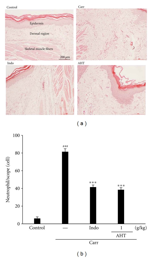
Figure 6.

Histological appearance of the mouse hind footpad after a subcutaneous injection with Carr stained with H&E stain at the 5th hr to reveal hemorrhage, edema, and inflammatory cell infiltration in control mice, Carr-treated mice demonstrating hemorrhage with moderately extravascular red blood cells and a large amount of inflammatory leukocyte mainly neutrophils infiltration in the subdermis interstitial tissue of mice, and mice given Indo (10 mg/kg) before Carr. AHT (1.0 g/kg) significantly shows morphological alterations (100x) (a) and the numbers of neutrophils in each scope (400x) compared to subcutaneous injection of Carr only (b). ### P < 0.001 as compared with the control group. ***P < 0.001 compared with the Carr group. Scale bar = 100 μm (one-way ANOVA followed by Scheffe's multiple range test).
