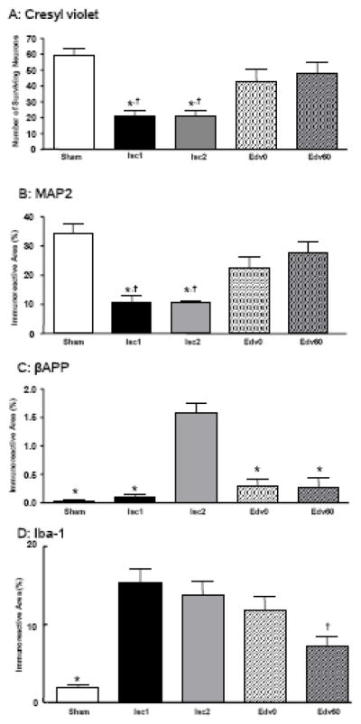
Fig. 3. Quantified Analyses of neuronal and axonal damages in the hippocampal CA1 region following global cerebral ischemia caused by cardiac arrest and resuscitation.

A: The extent of neuronal perikaryal damage is quantified by counting the number of surviving neurons in a predetermined hippocampal CA1 region. * P<0.05 compared with the Sham group; † P<0.05 compared with the Edv60 group. B: Neuronal perikaryal damage, quantified by the percentage of MAP2 immunoreactive areas in a predetermined hippocampal CA1 region. * P<0.05 compared with the Sham group; † P<0.05 compared with the Edv60 group. C: Axonal damage, quantified by the percentage of the βAPP immunoreactive areas in a predetermined CA1. * P<0.05 compared with the Isc2 group. D: Microglial activation, quantified by the percentage of the Iba-1 immunoreactive areas in a predetermined hippocampal CA1 region. * P<0.05 compared with the Isc1 and Isc2 groups; † P<0.05 compared with the Isc1 and Isc2 groups.
