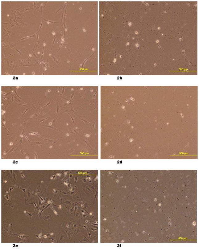
Fig. 2.
(2a – 2f) Light microscopic images of fetal rat neuronal cells, which underwent 96 hours of hypoxic stress and re-oxygenation with resuscitation by co-culturing with rat umbilical cord matrix derived stem cells on Day 2 (2a), Day 3 (2c) and Day 5 (2e). The size and morphology of the neuronal cells are distinctive from the larger stem cells. The control groups after the hypoxic stress and re-oxygenation were also shown on Day 2 (2b), Day 3 (2d), and Day 5 (2f).

