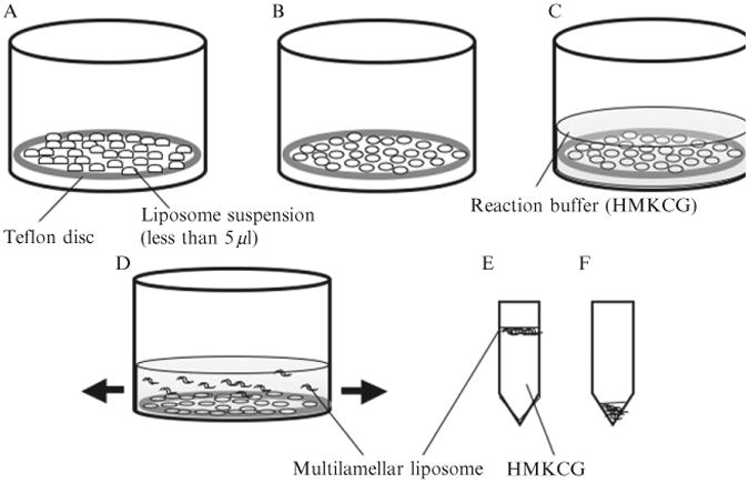
Figure 1.1.

Schematic illustration of the production of multilamellar liposomes. (A) A 37 mm Teflon disc is placed in a small beaker and 250 μl total aqueous suspension of lipid is deposited as many small drops (each drop is less than 5 μl). (B) The drops are dried with an air current and (C) the Teflon disc is covered with 5 ml of HMKCG and incubated overnight at 37 °C. (D) The Teflon disc is gently agitated and multilamellar liposomes floated off. (E) One microliter of the most concentrated suspension, nearest the Teflon, is transferred to an Eppendorf tube and left on the bench for 1 h. The liposomes rise to the surface. (F) HMKCG is carefully removed from the bottom, leaving a concentrated suspension of liposomes.
