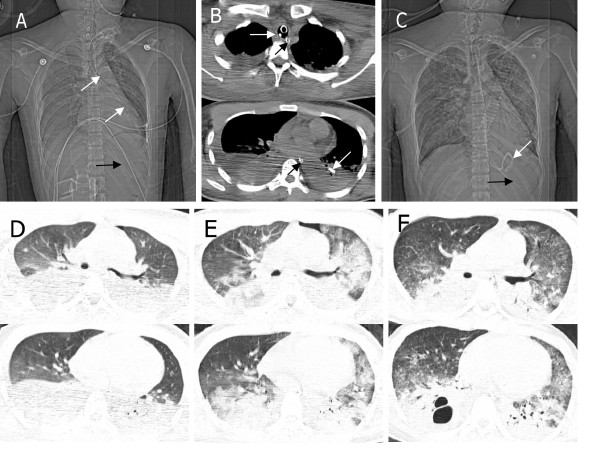
Figure 1.
Chest radiological images in the sixth patient with tracheobronchial malposition of the feeding tube. (a,b) Chest computed tomography (CT) on the first day showed the enteral feeding tube (white arrows) was misplaced into the left tracheobronchial tree and a nasogastric tube (black arrow) for drainage of gastric content was in the esophagus and the stomach. (c) Chest radiography on the second day showed the tip of the enteral feeding tube (white arrow) was in the stomach. Four signs indicate the right position: 1) the tube path follows the esophagus; 2) the tube bisects the carina; 3) the tube crosses the diaphragm in the middle; 4) the tip is below the left hemi-diaphragm. (d) Chest CT on the first day showed pulmonary contusion, pleural effusion and atelectasis. (e) Progression of bilateral bronchopneumonia was apparent on day 3. (f) Improvement of bronchopneumonia was apparent on day 7.

