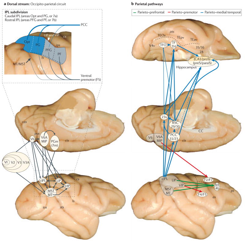
Figure 2. Anatomy of the three pathways.
a | The complexity of the occipito–parietal connections (shown by black arrows) on standard medial and lateral views of a rhesus monkey brain. The parts of visual area 1 (V1; also known as the primary visual cortex) that represent the central as well as peripheral visual fields are strongly connected with middle temporal area (MT) through visual areas V2, V3 and V4. By contrast, the parts of V1 that represent both the central and peripheral visual fields project through visual areas V2, V3 and V3A to a retinotopically organized and functionally distinct area V6 on the rostral bank of the parieto-occipital sulcus (pos). The visual information from area V6 reaches the parietal lobe through two main channels: one projecting medially to the bimodal (visual and somatosensory) area V6A and medial intraparietal area (MIP), which are located on the rostral bank of pos and the medial bank of the caudal intraparietal sulcus (ips), respectively; and the other projecting laterally to lateral intraparietal area (LIP) and ventral intraparietal area (VIP) in the ips and to areas MT and MST in the caudal superior temporal sulcus (sts). All of these posterior parietal areas are strongly connected with each other and with the surface cortex of the inferior parietal lobule (IPL). Feed-forward projections from lower- to higher-level processing areas (shown by single-ended arrows in the main figure) are usually reciprocated by feedback projections (not shown) from higher to lower areas; connections between areas at the same hierarchical level are shown by double-ended arrows in the main figure. The inset, which illustrates the findings from a study by Rozzi et al.19, depicts connections of IPL subdivisions with the posterior cingulate cortex (PCC) and ventral premotor area F5. Note that areas Opt and PG (subdivisions of the caudal IPL (cIPL)), which are primarily visual, appear to have stronger reciprocal connections with the PCC than with F5 (thick versus thin lines). The reverse holds for areas PFG and PF (subdivisions of the rostral IPL (rIPL)), which are primarily somatosensory. b | The sources and targets of the three pathways that emerge from the parietal component of the occipito–parietal circuit (also known as the dorsal stream). The parieto–prefrontal pathway (shown by green arrows) links areas LIP, VIP and MT/MST with a pre-arcuate region (area 8A; the frontal eye-field) and the caudal part of the principal sulcus in the lateral prefrontal cortex (area 46) — targets that serve eye movement control and spatial working memory, respectively. The parieto–premotor pathway (shown by red arrows) links areas V6A and MIP with the dorsal premotor cortex (areas F2 and F7), and also links area VIP with the ventral premotor cortex (areas F4 and F5) — targets that serve visually guided eye movements, reaching and grasping. The parieto–medial temporal pathway (shown by blue arrows; thick, thin and dashed lines represent dense, moderate and light projections, respectively) originates in the cIPL — that is, areas Opt and PG (see part a, inset) — and projects to subdivisions of the hippocampus (CA1/prosubiculum (proS), and presubiculum (preS)/parasubiculum (paraS)), both directly and indirectly via the PCC (areas 31 and 23), retrosplenial cortex (RSC; areas 29 and 30) and the posterior parahippocampal cortex (areas TF and TH in the rostral portion, and area TFO in the caudal portion) — targets that enable navigation and route learning. 23v, ventral subregion of the posterior cingulate; 28, entorhinal cortex; 35 and 36, perirhinal cortex; as, arcuate sulcus; cas, calcarine sulcus; CC, corpus callosum; cis, cingulate sulcus; cs, central sulcus; ios, inferior occipital sulcus; ls, lateral sulcus; ots, occipitotemporal sulcus; PGm, medial parietal area (also known as 7m); ps, principal sulcus; TE, rostral inferior temporal cortex; TEav, anterior ventral subregion of TE; TEOv, ventral subregion of TEO; TEpv, posterior ventral subregion of TE.

