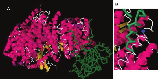
Figure 3.

A. Crystal structure of CRM1, snurportin1 and RanGTP. The heat repeat anti-parallel alpha helices are in red, RanGTP is in yellow, and snurportin is in green. B. A portion of the crystal structure image of CRM1, snurportin1 and RanGTP highlighting the alpha helix containing Cys-528 and the hydrophobic cleft that binds a cargo protein’s NES domain. The blue circle marks the position of the Cys-528 residue. The green backbone represents the amino terminal end of SNP1 which contains the NES domain of the protein. Image adapted from the RCSB PDB (www.pdb.org) of PDB ID 3GJX [50].
