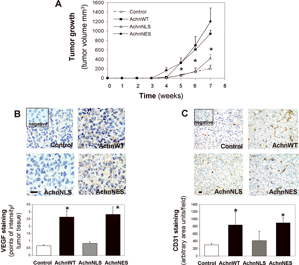
Figure 6. Nuclear Achn exacerbates tumor growth in vivo.
MDA-MB-231 cells expressing ectopic Achn-GFP fusion proteins were injected into immunodeficient SCID mice as described in the Methods. Each group contained six mice. A.. Tumor sizes were measured weekly for 7 weeks and growth curves plotted. (Mean ± SE, n=6). * = P<0.05 compared with the control group at the same time point. B. Paraffin-embedded tumor sections were processed for VEGF staining with IHC in which brown staining indicated positive signal. The images represented one of six tumor samples in each group and the data were analyzed using intensity quantification as described in the Methods. C. Frozen tumor samples were analyzed for vessel density using IHC staining of CD31. A vessel area with positive CD31 staining in 6–8 fields of each section was calculated using the NIH Image J analysis program. Mean ± SE, n=5–6. *=P<0.05 compared with control or AchnNLS. Bars=10 µm.

