
Figure 1. High-throughput screening of biomaterials for clonal growth.
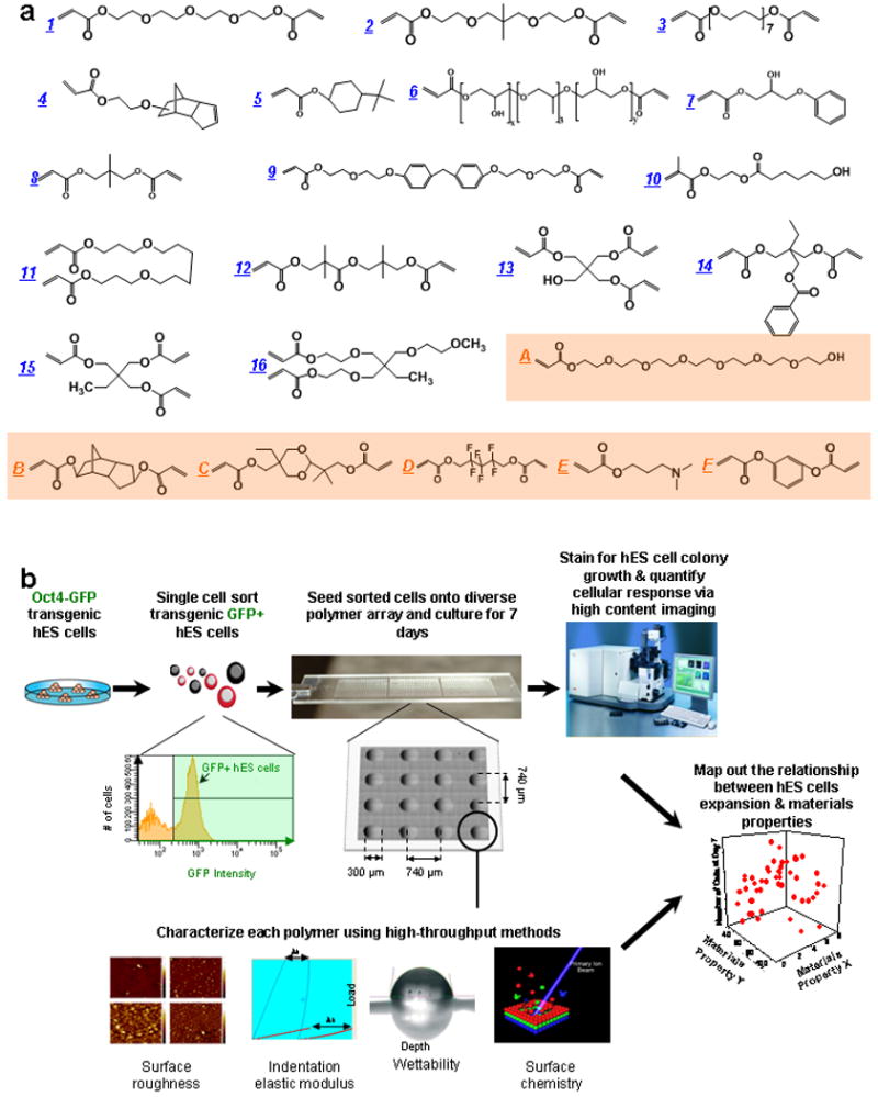
a, Monomers used for array synthesis were classified into two categories: “major” monomers that constitute >50% of the reactant mixture and “minor” monomers that constitute <50% of the mixture. Sixteen major monomers were named numerically (blue), and six minor monomers were labeled alphabetically (orange). b, Schematic of screen. First, transgenic Oct4-GFP hES cells were maintained on mEFs. Then flow cytometry enabled the isolation of high purity undifferentiated hES cells from the completely dissociated coculture of hES cells and mEFs. A flow cytometry histogram during a representative cell sort is shown. GFP+ cells (right of the black gate) were seeded onto the arrays, while the differentiated cells and mEFs (GFP-, left of the black gate) were not utilized. A photograph of the polymer microarray with 16 polymer spots is shown to illustrate dimensions and separation. Each polymer was also characterized using high-throughput methods to characterize its surface roughness, indentation elastic modulus, wettability (water contact angle, θC) and surface chemistry. Finally, cellular response on polymer array was quantified by using laser-scanning cytometry, and structure-function relationships were determined by numerical analysis of both the cellular response and materials characterization data.