
Figure 2. Diverse hES cell behavior on primary polymer arrays.
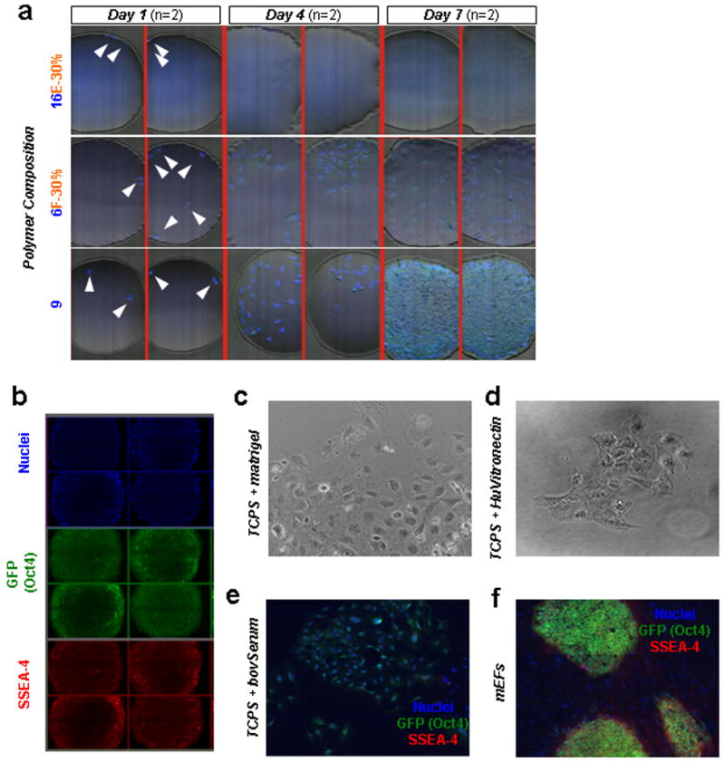
a, Single Oct4-GFP+ hES cells were seeded on the polymer arrays. White arrowheads point to cells attached after one day of culture, indicating a near clonal seeding density for each spot. Diverse cell behavior was seen on the array upon subsequent culture in mEF-conditioned media. Representative images of cell nuclei (stained by Hoechst in blue) on three different polymers (shown are two replicates of each): the 16E-30% polymer did not support either attachment or survival of dissociated hES cells; the 6F-30% supported moderate growth but also differentiation of hES cells; the 9 homopolymer (a “hit” polymer) supported robust growth of hES cells. b, Immunostaining of hES cells propagated on “hit” polymer spots for cell nuclei (blue) and for pluripotency markers Oct4 (green) and SSEA4 (red). Due to the raised center of each spot above the plane of the microscope slide, spot centers are not completely in focus, leading to lower intensity at the center of each image. c-e, At the near clonal cell densities used for the polymer experiments, hES cells spread out on matrigel-coated tissue culture polystyrene (TCPS), vitronectin-coated TCPS, and bovine serum-coated TCPS substrates in mEF-conditioned media. f, In contrast, traditional means of culturing hES cells by using mitotically-inactivated mouse embryonic feeder cells grown on gelatin-coated TCPS (“MEF substrate”) could support colony formation at these near clonal cell densities. In e and f, immunostaining was performed for nuclei (blue) and for pluripotency markers Oct4 (green) and SSEA-4 (red). See also Figure 6a for colony formation efficiencies.