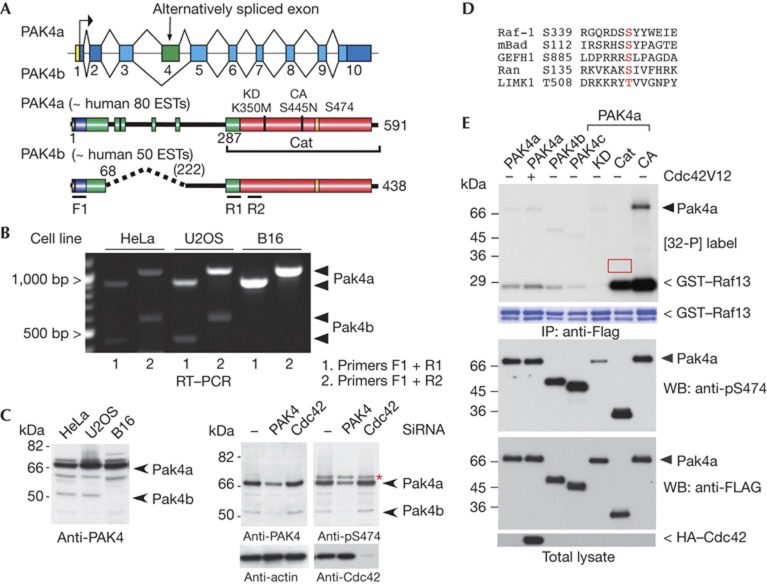
Figure 1.
PAK4 activity is independent of Ser 474 phosphorylation. (A) Schematic of the PAK4 gene in chromosome 19q13.2 and the exon organization of human PAK4. The PAK4b splice isoform lacks exon 4: identified domains include the nuclear localization signal (NLS, black), Cdc42–Rac interaction/binding (CRIB, blue) and catalytic domain (red). Other conserved but undefined regions in group II PAKs are in green. The number of human EST (expressed sequence tags) in the NCBI database is indicated. (B) Reverse transcriptase–PCR analysis (RT–PCR, 30 cycles) of mRNA isolated from HeLa, U2OS and mouse B16 cell lines. The position of the PAK4a and PAK4b cDNA products are marked. (C) Western blots (WBs) of cell lysates derived cell lines (40 μg) as indicated were probed with affinity-purified antibodies raised against PAK4(1–280) or a pSer 474 13mer peptide. The PAK4 and Cdc42 targeted short interfering RNAs (siRNAs) have been described previously [12]. The asterisk marks a 72-kDa nonspecific band. (D) Alignment of sequences surrounding known PAK4 substrate sites. (E) Flag–PAK4 constructs were immunoprecipitated (IP) and assayed in vitro with [32-P]ATP with the Raf1 S338/9 peptide substrate (see Methods). Autoradiograph on the top panel is a 2-h exposure. The A-loop phosphorylation was probed using CS#3241. The red box highlights that PAK4–Cat does not undergo auto-phosphorylation. CA, constitutively active, PAK4(S445N); Cat, PAK4(286–591); GST, glutathione S-transferase; HA, haemagglutinin; KD, kinase dead, PAK4(K350M); PAK, p21-activated kinase; PAK4c lacks residues 121–285, under accession number AAH02921.1.

