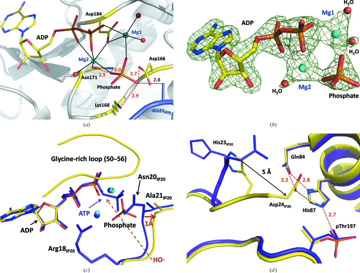
Figure 3.
(a) Active site of PKAc in the room-temperature structure RT PKA–Mg2ADP·PO4–IP20. Mg2+ coordination with the enzyme residues and water molecules is shown as black solid lines. (b) Electron density for ADP, phosphate, Mg2+ and water molecules in RT PKA–Mg2ADP·PO4–IP20 contoured at the 1.4σ level. (c, d) Superposition of the room- and low-temperature structures RT PKA–Mg2ADP·PO4–IP20 (yellow C atoms, cyan Mg2+ ions) and LT PKA–Mg2ATP–IP20 (purple C atoms and Mg2+ ions), respectively. Hydrogen bonds are shown as orange dashed lines. C—H⋯O interactions are shown as brown dashed lines. All distances are in Å.

