Fig. 1.
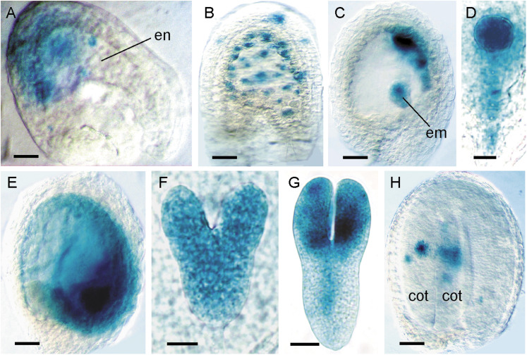
Expression analysis of pCYCB1;1:CYCB1;1DB-GUS during seed development. In all panels, seeds are oriented with the chalazal pole to the left and the micropylar pole to the right. (A) Fertilized ovule with GUS activity localized to dividing nuclei of an early syncytial endosperm (en). (B) Late syncytial endosperm-stage seed containing globular-stage embryo with GUS staining in dividing endosperm nuclei and integuments. (C) Late syncytial-stage seed with expression in the peripheral endosperm domain and embryo (em). (D) Globular-stage embryo with uniform GUS activity in the embryo proper and suspensor. (E) Early cellularized endosperm-stage seed containing heart-stage embryo with strong expression in micropylar endosperm and embryo. (F) Uniform GUS expression in a heart-stage embryo. (G) Torpedo-stage embryo with GUS activity in the cotyledons, shoot apex and provascular tissue. (H) Mature embryo seed stage with GUS staining restricted to dividing cells of the cotyledons (cot). Bars = 20 μm (A, F), 50 μm (B, C), 12 μm (D), 100 μm (E, H), 25 μm (G).

