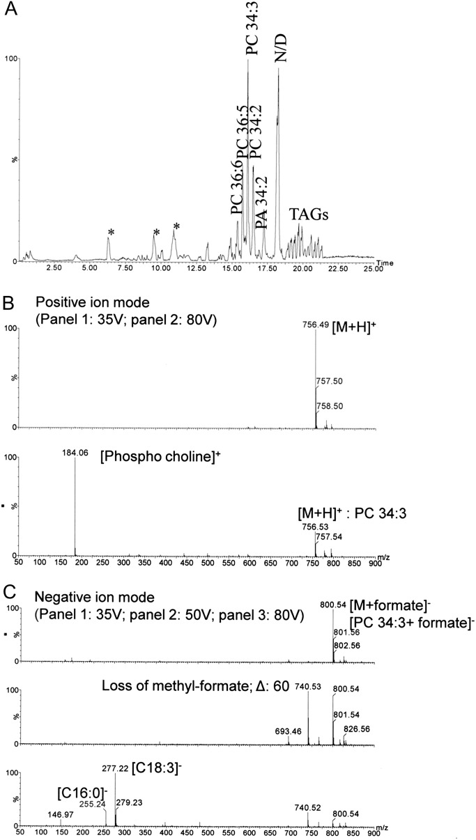
Fig. 5.
(A) Lipid profile of phloem exudates of Arabidopsis (chloroform phase) using LC-MS/positive ion mode. Mass spectra of the lipid at a retention time of 16.1 min were generated at aperture 1 voltages of 35, 50, and 80 V in both positive (B) and negative (C) ion mode to show lipid fragmentation. PC, phosphatidylcholine; PA, phosphatidic acid; TAG, triacyl glycerol; *, detergent; N/D, not determined due to multiple peaks in the spectra. Chromatograms are representatives of three biological replicates for each sample.

