
Figure 4.
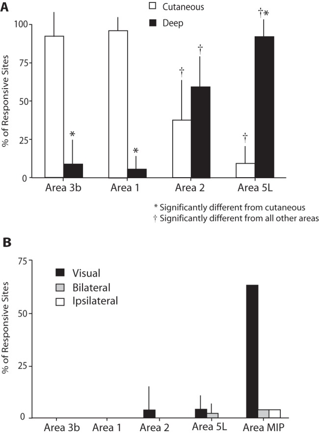
Histograms depicting the percentage of recording sites in which neurons in anterior and posterior parietal areas responded to (A) cutaneous versus deep stimulation or (B) visual, bilateral, or ipsilateral stimulation. The submodality to which neurons respond is distinct across cortical areas. The responsiveness of neurons to visual stimulation in area MIP is one of the distinguishing features of this field. Mean + standard deviation, P < 0.05. *—significantly different from cutaneous. †—significantly different from all other areas.