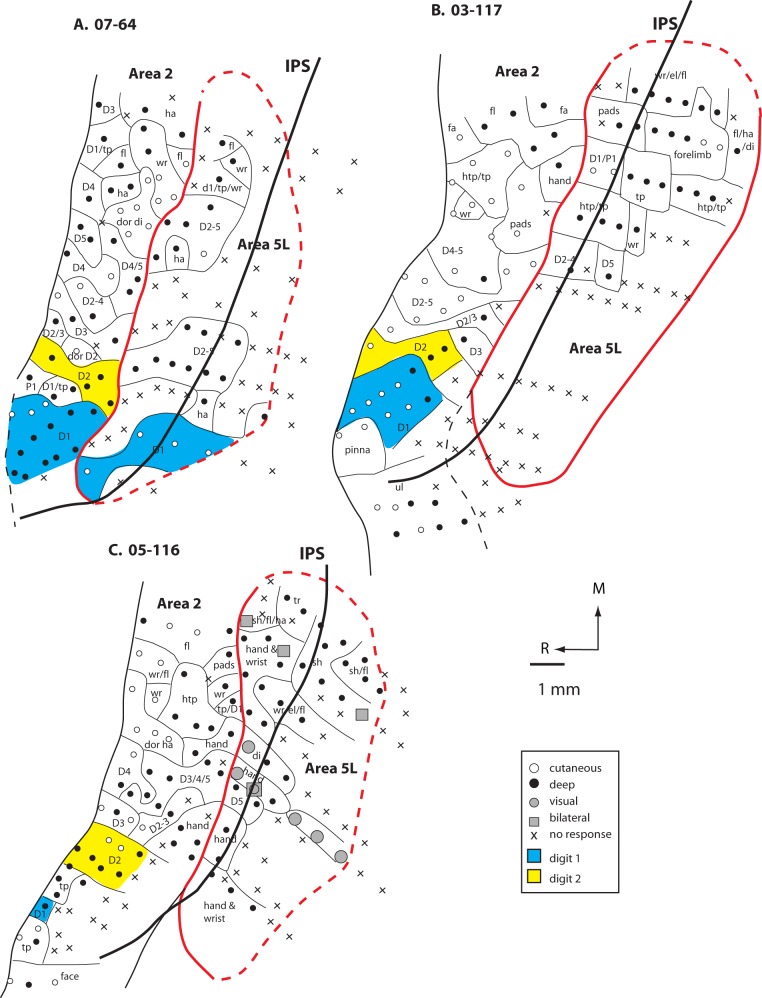
Figure 6.
Maps of area 5L and portions of area 2 generated from densely spaced electrophysiological recordings in 3 monkeys (A. 07-64, B. 03-117, and C. 05-116). In these cases, about half of the neurons in area 5L were responsive to stimulation of deep receptors of the skin, muscle, and joints (closed circles) and about half of the sites were unresponsive to any type of stimulation (black X). In one case, there were a few sites in which neurons responded to visual stimulation and 4 sites in which neurons had bilateral receptive fields (C). The topographic organization of area 2 could be readily discerned, but in area 5, the same body part was represented multiple times and was included in many different receptive field configurations. Furthermore, only the forelimb was represented in area 5L. Conventions as in previous figures.

