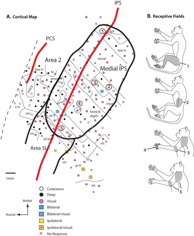
Figure 8.
A map of the medial half of the rostral bank of the IPS (A) and the location of the recording sites relative to the CS and IPS. Unlike area 5L, this medial IPS region contains numerous sites where neurons are responsive to visual stimulation and more sites in which receptive fields were ipsilateral or bilateral. (B) Examination of receptive field progression demonstrates that as recording sites move from medial to lateral (sites 1–5 in A), corresponding receptive fields progress from hindlimb, trunk, to forelimb. Thus, the entire body appears to be represented in the medial region. However, like area 5L, the same body part can be represented multiple times in different locations. Conventions as in previous figures.

