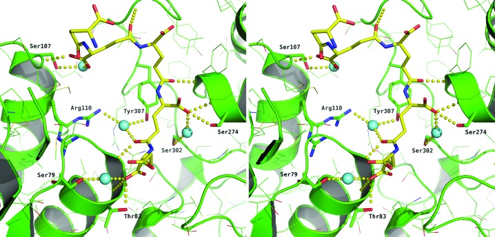
Figure 3.
Stereoview of the binding of PGA-A to LmCS. PGA is shown as in Fig. 1 ▶ and LmCS is shown in green, with N and O atoms of specific side chains coloured blue and red, respectively. Water molecules are shown as cyan spheres and hydrogen-bonding interactions are depicted as yellow dotted lines.

