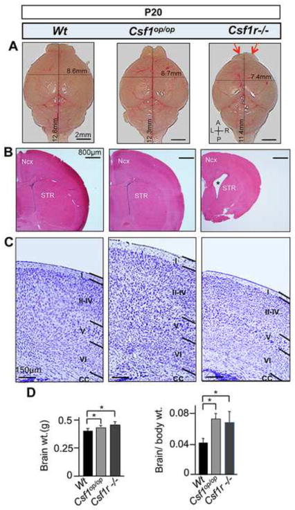
Fig. 1.
Gross anatomical and histological alterations in P20 Csf1op/op and Csf1r−/− brains. (A) A graded reduction in brain size along the A/P axis, from Csf1op/op to Csf1r−/− mice, with specific atrophy of the OB (red arrows) and a reduction in brain size along the mediolateral (L/R) axis in the Csf1r−/− mice (upper panels). (B) Coronal sections stained with hematoxylin and eosin (H&E), showing normal patterning but decrease in forebrain size of the Csf1r−/− mice. Asterisk indicates the increased size of the lateral ventricles in Csf1r−/− mice. (C) Nissl staining showing a normal laminar patterns, but an increase in thickness of neocortex (layers I–IV) in Csf1op/op mice and a reduction in thickness of the Csf1r−/− neocortex (all layers). (D) Whole brain weights (left panel) and brain to body weight ratios (right panel). *, p<0.01. n ≥ 5 per condition and mutant model. Ncx, neocortex; STR, striatum; A/P anterio-posterior; L/R, left-right; CC, corpus callosum; OB, olfactory bulb.

