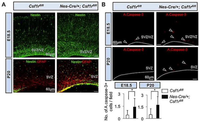
Fig. 8.
Expansion of forebrain progenitor pools and enhanced cellular apoptosis in Nes-Cre/+; Csf1rrfl/fl mice. (A) Immunofluorescence microscopy of coronal sections of E18.5 (upper panels) and P20 (lower panels) Wt and mutant neocortex. Upper panels, Nestin. Lower panels, Nestin-GFAP double immunostaining. (B) Photomicrographs of active caspase-3+ (red) apoptotic cells in the SVZ/VZ region of E18.5 (upper panels) and SVZ region of P20 (middle panels) Wt and mutant mice. Arrowheads indicate apoptotic cells. Dotted lines in (B) delineate the ventricular border. Lower panels: Quantitation of the number of active caspase-3+ apoptotic cells/field. Means ± SD of eight representative low-power (20X) fields per region per genotype from two different mice per genotype; *, P<0.01.

