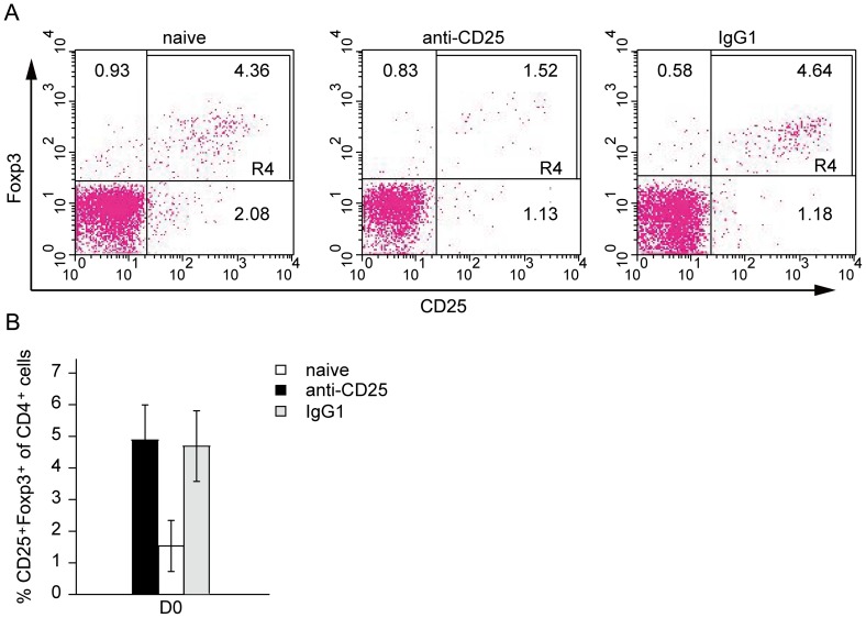
Figure 1. Effective depletion of CD4+CD25+ T cells in mice treated with anti-CD25 antibody.
C57BL/6 mice (6 per group) were intraperitoneally administered anti-CD25 mAb or rat isotype control IgG1 in 500 µg/mouse on day -3. Peripheral blood was obtained by retro-orbital bleed, red blood cells were excluded on day 0, and samples were then subjected to flow cytometry analysis for CD3, CD4, CD25 and Foxp3. (A) Representative flow cytometry data indicating the percentages of CD4+CD25+Foxp3+ Tregs in the peripheral blood of mice treated with anti-CD25 mAb. Double-staining for CD25 and Foxp3 expression in cells gated for CD3+ and CD4+. Values indicate the percentage of events in the indicated quadrant. (B) Bar graph depicting the percentages of CD4+CD25+Foxp3+ Tregs isolated from the peripheral blood of mice treated with anti-CD25 or isotype IgG1. The data are expressed as the mean values of two experiments with three mice per group.

