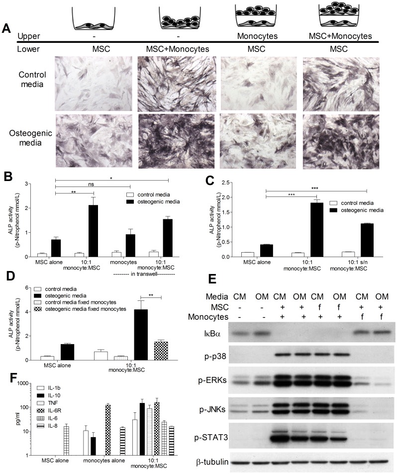
Figure 2. Osteogenic induction requires cell contact and monocyte-derived soluble factors.
MSCs were cultured either alone, with 10∶1 monocytes in contact, with monocytes separated by a 0.4 µm pore size cell culture insert (upper chamber) or with a co-culture of 10∶1 monocyte:MSC in the cell culture insert (upper chamber), in the presence or absence of osteogenic stimuli. After 7 days, MSC ALP was assessed by staining (A) and quantification of enzyme activity (B). ALP activity induced by 10∶1 monocyte:MSC co-cultures was compared to that induced by treatment with supernatant from co-cultures incubated for 24 hours (10∶1 s/n) (C). ALP activity of MSCs was also measured after 7 days of co-culture with either fresh of fixed monocytes (fixed in 0.05% glutaraldehyde for 1 min followed by 1 min in 0.2 M glycine) (D). MSC were treated for 30 mins with supernatant from monocyte:MSC co-cultures or monocyte:MSC co-cultures where either monocytes or MSCs had been fixed (f), in control media (CM) or in osteogenic media (OM) and then lysed (E). MSCs alone in CM and OM were used as controls. MSC lysates (10 µg per condition) were subjected to WB for detection of IκBα, p-p38, pERKs, p-JNKs and pSTAT3. Anti-β-Tubulin was used as a loading control. The levels of IL-1β, IL-10, TNFα, IL-6, IL6R and IL-8 were measured in culture media from MSC or monocyte alone cultures or 10∶1 monocyte:MSC co-cultures (F). Phase contrast pictures (10X) are representative of three independent experiments performed. Graphs show means ± SEM of at least three independent experiments performed in triplicate. *p≤0.05, **p≤0.01, ***p≤0.001 Blots are representative of three independent experiments performed.

