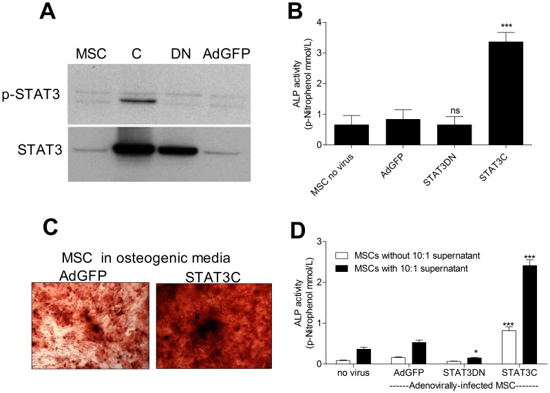
Figure 3. OSM mediates monocyte osteogenic effect through STAT3 signaling.
The levels of pSTAT3 and STAT3 were assessed using WB in MSCs infected either with STAT3C (50 M.O.I.) or STAT3DN (100 M.O.I.) adenoviruses in osteogenic media. GFP adenoviral infection was used as a control (100 M.O.I.) (A). MSC ALP activity was measured 7 days after infection either with STAT3DN, STAT3C or AdGFP (B). MSC infected with the STAT3C or AdGFP viruses were also kept for 21 days in osteogenic media when bone nodule formation was assessed using Alizarin Red S staining (C). MSC infected with either the STAT3DN, STAT3C or AdGFP were kept with or without conditioned supernatant from separate monocyte:MSC co-cultures (10∶1 s/n) for 7 days when ALP activity was quantified (D). Blots are representative of three independent experiments performed. Graphs show means ± SEM of three independent experiments performed in triplicate. Phase contrast pictures (10X) are representative of three independent experiments performed. *p≤0.05, ***p≤0.001.

