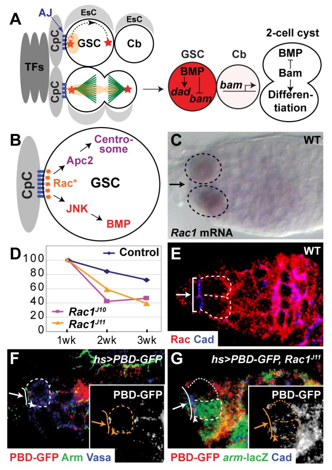
Figure 1. Activated Rac is localized to the CpC-GSC interface.

(A) GSCs in the CpC niche. Two to three GSCs are present in a niche composed of Terminal Filament cells (TFs), Cap Cells (CpCs), and Escort Cells (EsCs). The GSCs are anchored to the CpCs by Adherens Junctions (AJs). Each GSC contains a cytoskeletal organelle, the spectrosome (light orange) adjacent to the niche. One centrosome (red star) is present at the niche-GSC interface. After centrosome duplication, a centrosome migrates around the cortex (dotted line with arrow). The plane of GSC division is perpendicular to the niche-GSC interface. One daughter cell, the Cystoblast (Cb), is born outside of the niche and the other daughter cell remains in the niche as a GSC. During GSC division, the spectrosome elongates, and some spectrosomal material is segregated to the Cb. GSCs have high levels of BMP signaling, which leads to the transcription of dad and the repression of bam expression. Cbs have much lower levels of BMP signaling and transcribe bam. Bam functions to promote Cb differentiation and to inhibit BMP signaling. (B) Model for Rac function in GSCs: Rac is asymmetrically activated (Rac*) at the CpC-GSC interface to orient interphase centrosomes and to promote BMP signaling in GSCs. (C) Wild-type ovariole in situ hybridized with Rac1 anti-sense probe (purple). Some, but not all, GSCs expressed Rac transcript. (D) Percentage of germaria carrying marked wild-type or Rac mutant clones as a function of time. Wild type (blue diamond), Rac1J10 Rac2Δ MtlΔ/Rac1J11 Rac2Δ+ (purple rectangle), and Rac1J11 Rac2Δ MtlΔ/Rac1J11 Rac2Δ+ (yellow triangle). (E) Rac is localized to the cortex of wild-type GSCs. Bracket, the CpC-GSC interface as marked by anti-Cadherin staining. (F) After mild heat shock, a biosensor for activated Rac, PBD-GFP, is localized at the CpC-GSC interface in wild-type GSCs. (G) PBD-GFP is not localized to the CpC-GSC interface after mild-heat shock in a Rac mutant (Rac1J11 Rac2Δ MtlΔ/Rac1J11 Rac2Δ+) GSC (dotted outline), marked by the absence of lacZ expression, compared to normal localization at the CpC-GSC interface in a Rac1J11 Rac2Δ MtlΔ/+++ GSC (dashed outline). (C, E–G) Arrow, CpC niche; dashed or dotted outline, individual GSC. (F–G) Solid line, CpC-GSC interface; arrowhead, asymmetrically localized PBD-GFP in GSCs. Inset: white, anti-GFP staining.
