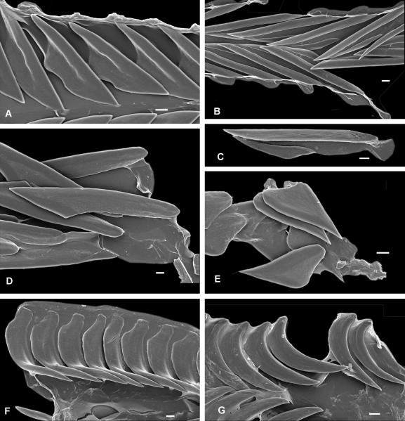
Figure 2.
Flat (A-E) and solid recurved (F-G) teeth of Terebridae. A – Pellifronia jungi (IM_2007_30591), ventral view of radular membrane, only half shown; B – Clathroterebra poppei (IM_2007_30546), ventral view of radular membrane; C – Terebra succincta (IM_2007_30582), separate marginal tooth; D – Terebra trismacaria (IM_2007_30579), ventral vies of radular membrane; E – Myurella lineaperlata (IM_2007_30635), group of teeth attached to the subradular membrane; F – Euterebra fuscolutea (IM_2009_10133), ventral view of radular membrane, only half shown; G – Duplicaria sp. 2 (IM_2009_10164), ventral view of radular membrane, only half shown. Scale bars – 10 μm.

