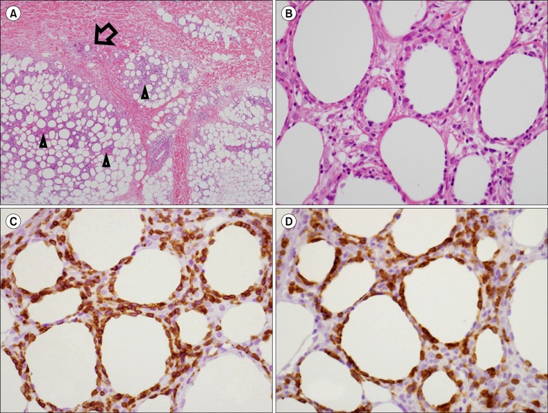
Fig. 1.
Biopsy of a skin nodule. (A) The skin tissue showed dense lymphocytic infiltrate in lobular panniculitis-like pattern (arrowheads) with focal dermal infiltrations (arrow) (hematoxylin and eosin stain, ×40 magnification). (B) The infiltrated lymphocytes showed atypical features with hyperchromatic, irregular nuclei, and occasional nucleoli. There was fat rimming with atypical lymphocytes (hematoxylin and eosin stain, ×400 magnification). (C) In immunohistochemical stain, the atypical lymphocytes were positive for CD3 (CD3, ×400) and (D) CD8 (CD8, ×400 magnification).

