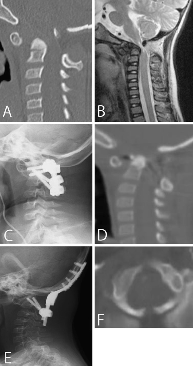
Fig. 2.

Case 6. A pre-operative CT scan reconstruction of the cervical spine (a) suggests an atlantoaxial subluxation, and a pre-operative sagittal MR image (b) demonstrates spinal canal stenosis and cord signal change. Postoperative radiograph (c) of the lateral cervical spine shows correction of atlantoaxial alignment using C1 lateral mass screw and C2 pedicle screws. 4 months after primary surgery, sagittal CT reconstruction of the cervical spine (d) shows stenosis between occipito and dens. After reoperation, radiograph of the lateral cervical spine (e) shows successful O-C2 fusion. Pre-operative axial CT scan of the atlas (f) shows spina bifida of the posterior arch
