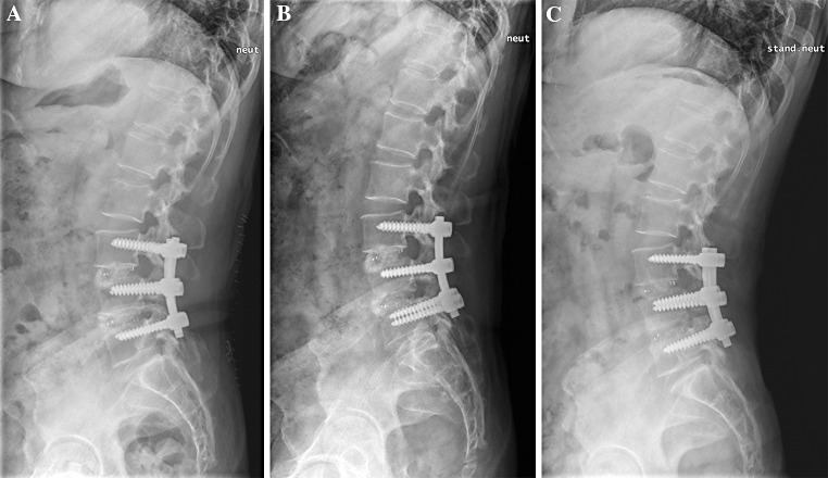
Fig. 1.
Lateral simple radiography of the 57 years of female patient with degenerative spondylolisthesis and spinal stenosis at L3-4-5 level, who undergone double level PLIF with porous hydroxyapatite bone chip in addition to local decompressed bone. a Immediate post-operative image showing clotty radio-opaque shadow of hydroxyapatite bone chips in interbody space. b 3 months after operation, irregular pattern of mixed radio-opaque and -lucent shadow is seen presumably due to uneven resorption of graft materials. c 12 months postoperatively, more organized shadow is observed representing trabecular bridging across interbody space

