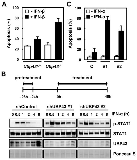
Fig. 1. Increased IFN-α/β-mediated apoptosis in UBP43-depleted cells.
(A) Bone marrow cells from Ubp43+/+ and Ubp43−/− mice were cultured in the absence or presence of IFN-β (500 units/ml) for 24 hrs, and apoptotic cell death was analyzed by FACS after staining with Annexin V-FITC. (B) THP-1 cells stably expressing control shRNA (shControl) or UBP43- specific shRNAs (#1 and #2) were treated with IFN-α, as indicated on the top of the panel. The cells were pretreated with 1,000 units/ml of IFN-α for 2 hrs, washed with regular medium without IFN-α, and cultured another 24 hrs. The cells were again treated with 1,000 units/ml of IFN-α and harvested at various time points. The total proteins were analyzed by immunoblotting with anti-STAT1, anti-phosphoSTAT1Tyr701 (p-STAT1), and anti-UBP43 antibodies. Relative protein loading was shown by Ponceau S staining. (C) Cells were treated with IFN-α as indicated in (B) and analyzed for apoptosis after 48 hrs of treatment using FACS. C, #1, and #2 represent the THP-1 cells stably expressing control shRNA, UBP43- specific shRNA#1, or UBP43-specific shRNA#2, respectively. All data in bar graphs throughout the figures represent the mean value for three independent experiments.

