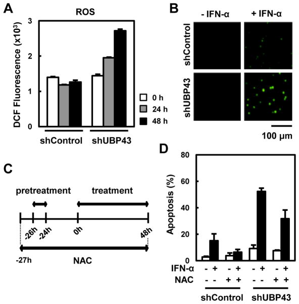
Fig. 4. Involvement of ROS in IFN-α/β-mediated apoptosis in UBP43-knockdown THP-1 cells.
(A) THP-1 cells stably expressing shControl or UBP43-specific shRNA#1 were treated with IFN-α as in Fig. 1B. The cells were harvested after the 2nd treatment of IFN-α at various time points, incubated with 10 μM H2DCFDA, and then analyzed by FACS. (B) The cells in (A) were harvested on a slide at 48 hrs after IFN-α treatment. An increase in ROS (green dots) was observed using a fluorescence microscope. (C and D) DMSO or NAC (10 mM) was added to the culture of THP-1 cells stably expressing shControl or UBP43-specific shRNA#1 one hour prior to IFN-α pretreatment as shown schematically in (C). The cells were harvested at 48 hrs after the IFN-α treatment and analyzed for apoptosis (D).

