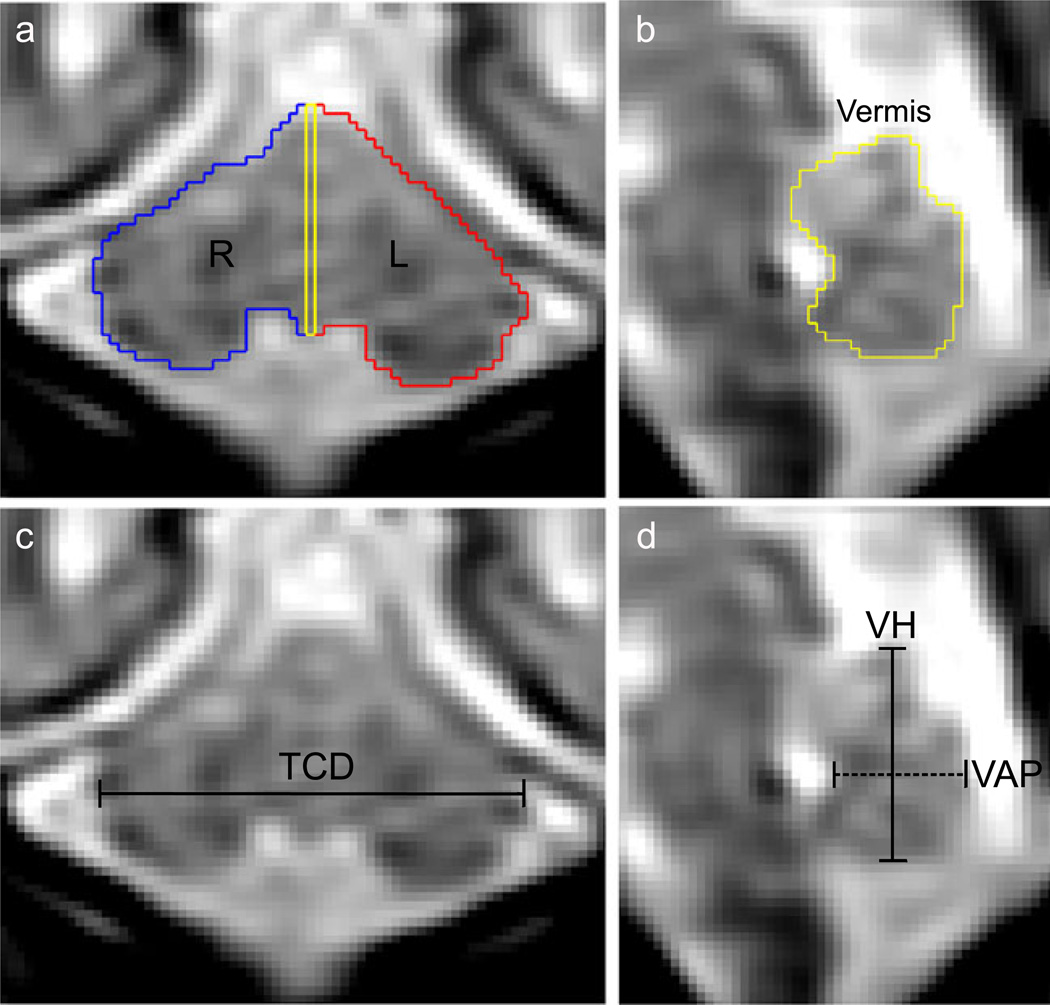
Fig. 2.
Example of cerebellum linear dimensions and volumetric segmentation at 25 gestational weeks. a, b) The perimeter of the cerebellum was manually drawn then labeled as right hemisphere (blue), left hemisphere (red), and midsagittal vermis (yellow). c Transverse cerebellar diameter (TCD) was measured at the maximum distance in the coronal plane. d Vermis height (VH; solid line) and anterior–posterior diameter (VAP; dashed line) were measured on the midsagittal slice Cerebellum

