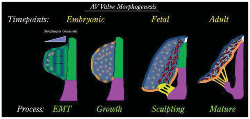
Figure 1. Schematic of AV Valve Development.

AV valve morphogenesis commences from an EMT event whereby morphogenic cues secreted by the AV junctional myocardium (green) diffuse through the cardiac jelly. Mesenchyme migrates towards these cues (arrows) in a directed fashion and undergoes a proliferative expansion to fill the cushion during the “growth phase”. As cells get closer to the myocardium (red cells), they change their secretory prolife and begin synthesizing fibrillar matrices such as collagens (black lines). During this fetal timepoint, cellular contraction and cell density facilitate valve sculpting. Development of the chordae tendineae (yellow) and the fibrous annulus (white) are markedly observed. The valve continues to attenuate and separate from the AV junctional myocardium (green), which is selectively removed yielding a tethered AV valve leaflet during adulthood.
