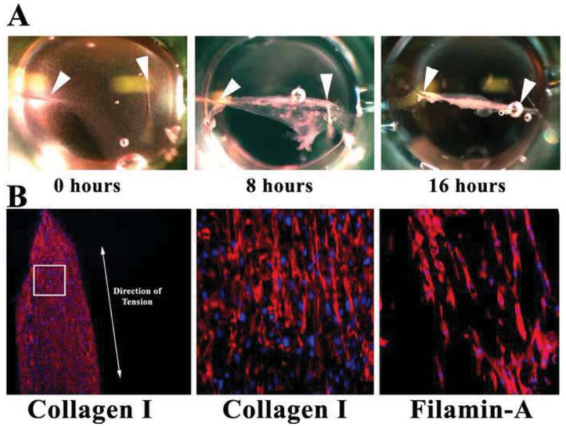
Figure 4. Fibrin gel assay.

(A) Pictures of fibrin gels compacting over time in 96-well dishes. Arrowheads demarcate where needles (“pillars”) were placed. Gel compacts to a linear construct in <1 day. (B) IHC of sectioned constructs showing de novocollagen I (red) and FLNA (red) align along the axis of tension (double-headed arrow). Nuclei-blue.
