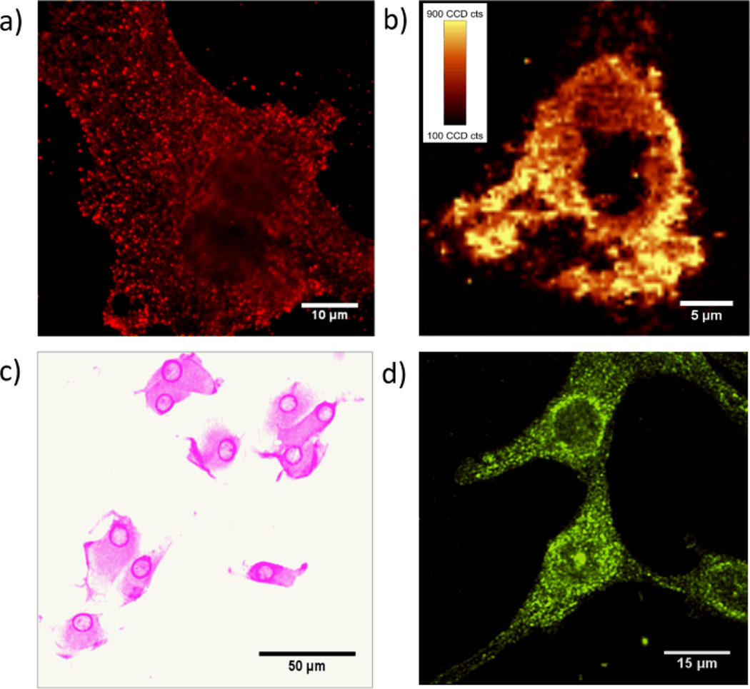
Figure 3.
MBA-pAuNPs-cRGD served as a multi-modality probe that enables αvβ3 integrin receptors on U87MG cancer cells to be imaged with four different imaging techniques at the single cell level. (a) Strong single-particle luminescence from pAuNPs enabled the integrin receptors on a single U87MG cell to be imaged using fluorescence microscopic imaging modality. (b) Raman image of a U87MG cancer cell labeled by cRGD-pAuNPs-MBA, which was constructed based on the Raman vibration of MBA at 1579±10 cm-1(fluorescence background was substracted). (c) Bright-field image of the cancer cells labeled by the NPs. Strong surface plasmons of the pAuNPs made the cells purplish. (d) Strong surface plasmon scattering of pAuNPs allow the cancer cells to be visualized using dark-field imaging modality.

