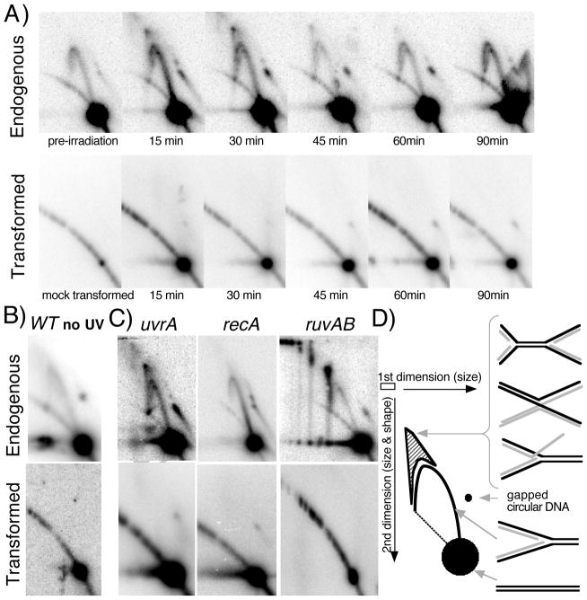
FIGURE 4. Replication- and RecA-dependent processing intermediates are not observed on newly introduced plasmids.
A) Replication-associated structural intermediates observed on endogenous plasmids containing UV-induced DNA damage are not observed on plasmids following transformation. Wild-type cells containing an endogenous plasmid pBR322 were UV-irradiated with 55 J/m2 (top) or wild-type cells were transformed with a plasmid that was UV-irradiated with 110 J/m2 (bottom). Genomic DNA was then purified, digested with PvuII, and analyzed by two-dimensional agarose-gel analysis at the times indicated. B) Non-damaged plasmids also fail to generate replication intermediates when first introduced into cells. Wild-type cells containing an endogenous plasmid pBR322 were mock UV-irradiated (top) or wild-type cells were transformed with a plasmid that was mock UV-irradiated, and then analyzed as in (A) following a 60-min recovery period. C) On endogenous plasmids containing DNA damage, RecA-dependent structural intermediates persist and accumulate in uvrA and ruvAB mutants (top), but are not observed when UV-irradiated plasmids are introduced into these mutants (bottom). uvrA, recA, and ruvAB mutants were analyzed as described in (A) following UV irradiation of cultures containing an endogenous plasmid or following introduction with UV-irradiated plasmid, pBR322. uvrA and recA mutants were analyzed at 30 min following treatment, ruvAB mutants were analyzed 60 min following treatment. D) Migration pattern of replication- and UV-induced structural intermediates following PvuII digestion of pBR322 during two-dimensional agarose-gel analysis. Nonreplicating plasmids run as a linear 4.4-kb fragment. Normal replicating fragments form Y-shaped structures and migrate more slowly because of their larger size and nonlinear shape, forming an arc that extends out from the linear fragment. Double Y- or X-shaped molecules migrate in the cone region and are observed after UV-induced damage on endogenous plasmids.

