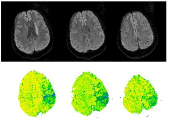
Figure 3.

DWI (top panel) and PWI (lower panel) scans of a patient with asyntactic comprehension and impaired short-term memory associated with ischemia in left angular gyrus. He was 40% correct for passive reversible sentences, but 85% correct for active reversible sentences. He was 85% correct for cleft-object sentences, and 100% correct for cleft-subject cleft sentences. Forward digit span of 3, and was unable to do backwards digits. The acute infarct on DWI involves only the caudate. PWI shows significant hypoperfusion (>4 sec. delay in TTP relative to the homologous regions in the right hemisphere; see methods) in angular gyrus (BA 39) and supramarginal gyrus (BA 40), but not Broca's area (BA 44 or 45).
