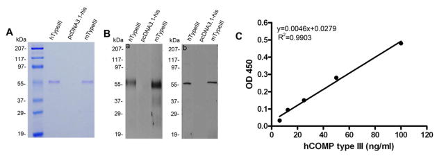
Figure 2. Characterization of recombinant COMP type III domain and the establishment of the sandwich ELISA using recombinant COMP type III as a standard.
(A) The HEK293-EBNA cells were transfected with either expression plasmid encoding His-tagged human COMP type III (lane 1), His-tagged mouse COMP type III (lane 3), or empty vector as a control (lane 2) and the conditioned media collected. Purified recombinant proteins using Ni-NTA resin was separated by non-reducing 10% SDS-PAGE gel and visualized with Coomassie Brilliant blue staining. (B) After purification with Ni-NTA resin, the expression of recombinant human COMP type III (lane 1) and mouse COMP type III (lane 3) was verified by western blot using anti-COMP pAb (a) or anti-COMP mAb2127F5 (b). No band was detected in the control media (lane 2). (C) The standard curve was generated by the serial concentration of diluted recombinant human COMP type III plotted against the OD450 value; the blank has been subtracted from all the absorbances.

