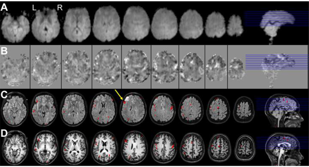
Figure 6.
Demonstration of the localization of Broca’s language area in a candidate for surgical resection of a brain tumor using PRESTO fMRI. Shown are 9 slices of a PRESTO experiment with 2D SENSE and 8-channel headcoil on a Philips 3T. Images are of language task (verb generation) in a patient with a left frontal glioma, used for surgical planning. Rows display: A - average PRESTO scan, B – T-maps of language activity (range −10 to 6), C – activity exceeding a threshold of t=5 (p<0.05 Bonferroni-corrected) projected onto a FLAIR scan, D – same on T1-weighted anatomical scan. The yellow arrow shows the location of Broca’s area in this subject (with the tumor in front). Scan parameters PRESTO: TR 22.5ms; TE 33.2ms; echo-shifting of 1 TR; flip angle = 10°; FOV 224×256×160mm3; matrix 56×64×40; voxel size 4.0mm isotropic; 0.6075 seconds per volume; 40 slices; sagittal orientation. Courtesy of Department of Neurosurgery, UMC Utrecht, The Netherlands.

