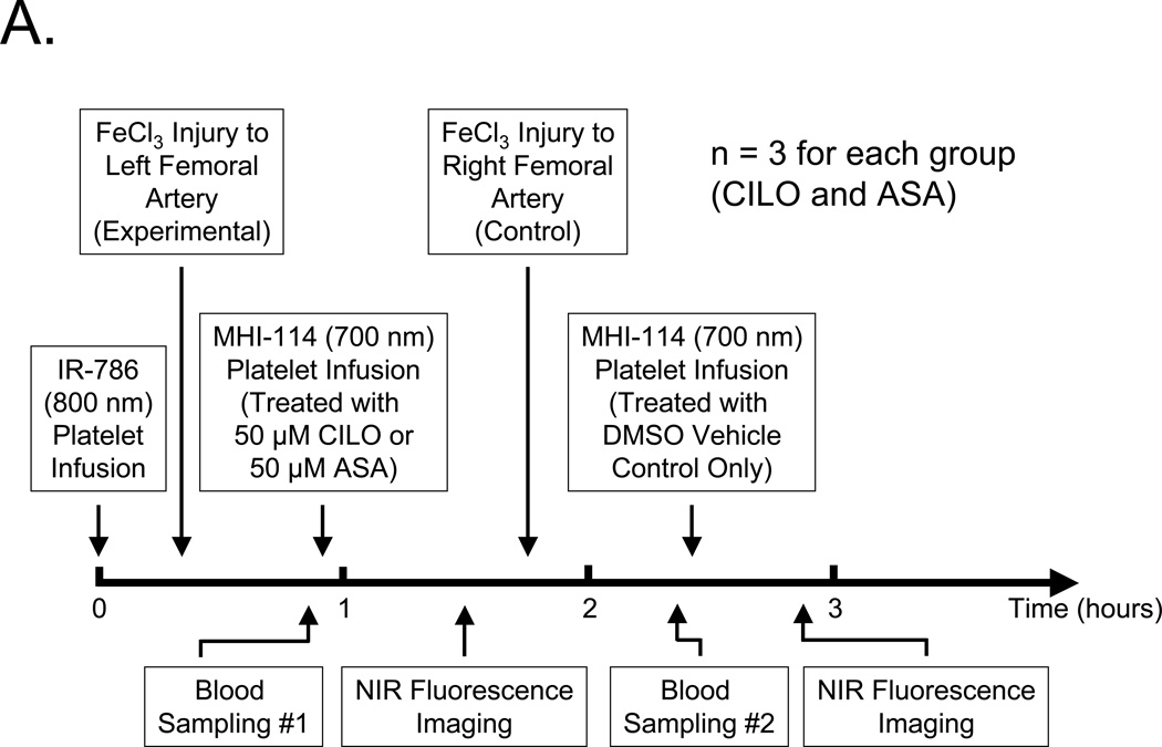
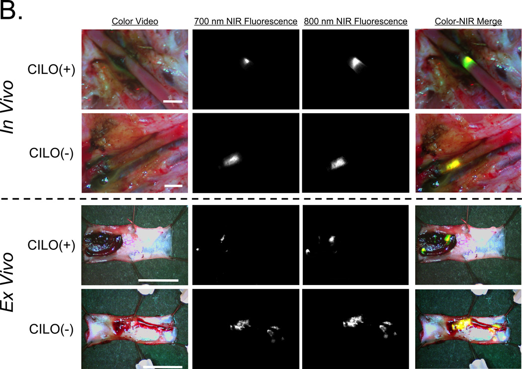
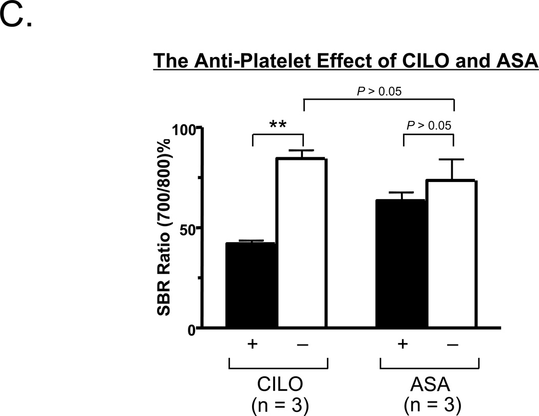
Figure 5. Quantitation of the effect of cilostazol (CILO) and aspirin (ASA) on platelet incorporation into preexisting thrombi.
A. Protocol for femoral artery injury, drug-treated platelet infusion, blood sampling, and intraoperative NIR fluorescence imaging.
B. Simultaneous 700-nm and 800-nm NIR fluorescence imaging of the intact femoral artery as described in detail in (A) after pretreatment with 50 µM CILO. Shown are color video, 700-nm NIR fluorescence, 800-nm NIR fluorescence, and a pseudo-colored merge of the 3, with red used as the pseudo-color for 700 nm, green for 800 nm, and yellow for co-localization in the merged image. Ex vivo surgical specimens are shown below the dashed line. Representative images are from n = 3 independent femoral artery thrombi using CILO-treated platelets. ASA results are not shown.
C. Comparison of the SBR ratios (700 nm/800 nm %; mean ± SEM) between CLIO (+) and CILO (−), or between ASA (+) and ASA (−) are shown. Shown are the P values for the indicated statistical comparisons by a one-way ANOVA followed by Tukey’s multiple comparisons test. ** = P < 0.01.


