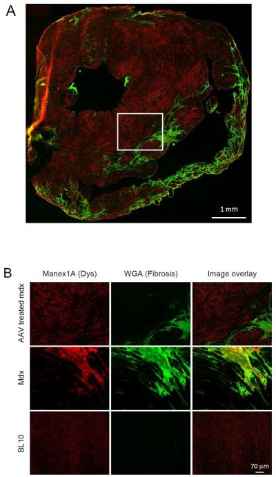
Figure 1. Evaluation of AAV micro-dystrophin transduction in the heart of >21-m-old mdx mice.
Myofibers that were successfully transduced by the AAV microgene vector showed bright red sarcolemmal staining. Fibrotic collagen deposition was revealed with Oregon green 488-conjugated wheat germ agglutinin (green). A, Representative immunofluorescence staining photomicrograph of the whole heart from a micro-dystrophin treated mdx mouse. B, Representative high power immunofluorescence staining photomicrographs of the heart from treated and untreated mdx mice, and normal BL10 mice. The images of AAV treated mdx mice correspond to the boxed area in panel A.

