Figure 3.
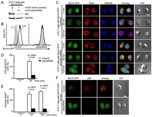
p62 silencing inhibits RelA nuclear translocation and blocks Bcl10 degradation. (A) D10 T cells expressing Bcl10-GFP plus either mir30 shRNA vector or mir30 p62shRNA were analyzed by immunoblotting to detect p62 expression. (B-E) D10 T cells expressing Bcl10-GFP plus mir30 shRNA vector or mir30 p62shRNA were stimulated with anti-CD3 for the indicated times. Cells were analyzed by flow cytometry to detect degradation of Bcl10-GFP (B); or stained with anti-RelA and the DNA dye, DRAQ5, and analyzed by confocal microscopy to assess RelA nuclear translocation (C). Graphs showing percentage of cells in (C) forming Bcl10-GFP clusters (POLKADOTS) (D) or with RelA nuclear translocation (E). (F) D10 T cells expressing Bcl10-GFP plus mir30 shRNA vector or mir30 p62shRNA were stimulated for 20 min with anti-CD3 and stained with anti-p62. p62 silencing and p62-Bcl10 co-localization were assessed by confocal microscopy. Means (+/− SEM) were calculated by counting at least 20 cells from each of three experiments. Scale bar = 10μm. Data are representative of two (A,B) or three (C,F) independent experiments. See also Figure S3.

