Figure 5.
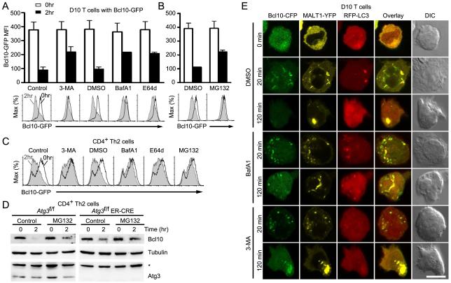
Blockade of autophagy prevents TCR-dependent Bcl10 degradation. D10 T cells (A,B) expressing Bcl10-GFP were pre-treated with distilled water (control), DMSO (vehicle control), the autophagy inhibitor 3-methyladenine (3-MA), or the lysosomal degradation inhibitors Bafilomycin A1 (BafA1) and E64d (A) or the proteasomal inhibitor MG132 (B), and stimulated for the indicated times with anti-CD3. Bcl10-GFP degradation was assessed by flow cytometry. Histograms are representative data from one experiment and bar graphs are mean (+/−SEM.) of Bcl10-GFP median fluorescence intensity (MFI) from three independent experiments. (C) The experiment of (A) and (B) was repeated using primary Th2 cells. (D) Primary Th2 cells expressing (Atg3f/f) or not expressing ATG3 (Atg3f/f ER-Cre) were pretreated with vehicle (control) or MG132, followed anti-CD3+anti-CD28 stimulation for the indicated times. Endogenous Bcl10, tubulin, and ATG3 were detected by immunoblotting. Asterisk indicates non-specific band. (E) D10 T cells expressing Bcl10-CFP, MALT1-YFP and RFP-LC3 were pre-treated with DMSO (control), BafA1, or 3-MA, followed by anti-CD3 stimulation. Cells were imaged by confocal microscopy to assess co-localization between proteins. Data are representative of two independent experiments. Scale bar = 10μm.

